Home » Without Label » Anatomy Diagram Rib Area / How Many Ribs Do Humans Have Men Women And Anatomy : We are pleased to provide you with the picture named heart, lung, diaphragm and ribs location.we hope this picture heart, lung, diaphragm and ribs location can help you study and research.
Anatomy Diagram Rib Area / How Many Ribs Do Humans Have Men Women And Anatomy : We are pleased to provide you with the picture named heart, lung, diaphragm and ribs location.we hope this picture heart, lung, diaphragm and ribs location can help you study and research.
Anatomy Diagram Rib Area / How Many Ribs Do Humans Have Men Women And Anatomy : We are pleased to provide you with the picture named heart, lung, diaphragm and ribs location.we hope this picture heart, lung, diaphragm and ribs location can help you study and research.. Related posts of anatomy of ribs and its related area abdominal artery anatomy. The bones of the rib cage are the sternum, the 12 thoracic vertebrae and the 12 pairs of ribs. Anatomy diagram rib area : Thoracic cavity description anatomy physiology britannica.ribs eight to ten are the false ribs and are connected to the sternum indirectly via the cartilage of learn everything about the ribs with our articles, video tutorials, quizzes, and labeled diagrams there are eleven pairs of external intercostal muscles and these are the most. The anatomy of the human ribs is made up of 24 ribs which are parted in 12 pairs (each on the left and right side of the chest wall), with the sternum, metasternum(the.
The rib cage is the arrangement of ribs attached to the vertebral column and sternum in the thorax of most vertebrates, that encloses and protects the vital organs such as the heart, lungs and great vessels. Human rib cage anatomy anterior and right lateral view all bones. The ribs partially enclose and protect the chest cavity, where many vital organs (including the heart and the lungs) are located. The ribs are a set of twelve paired bones which form the protective 'cage' of the thorax. Rib bones are not classified as long bones.instead, anatomists classify the ribs as flat bones, and they are located within the axial skeleton.together with the sternum, thoracic vertebrae, and costal cartilages, the ribs form the thoracic cage, also called the bony thorax.
The Thoracic Cage The Ribs And Sternum Human Anatomy And Physiology Lab Bsb 141 from s3-us-west-2.amazonaws.com Anatomy diagram rib area : The ribs are a set of twelve paired bones which form the protective 'cage' of the thorax. In this image, you will find thoracic vertebrum, costochondral joint, costal cartilage, costal margin, costal arch, thoracic vertebrum, xiphoid process, xiphisternal joint, body, manubrial sternal joint, manubrium, the sternal notch in it. Bony surface landmarks on the back note the area of the triangle of ribs anatomy types ossification clinical significance how to surface anatomy and skeleton of the thorax questions and study guide human rib cage anatomy diagram including anterior and right lateral But this number may be increased by the development of a cervical or lumbar rib, or may be diminished to eleven. It has a roughened area on its upper surface, from which the serratus anterior muscle originates. Abdominal artery anatomy 12 photos of the abdominal artery anatomy abdomen arteries anatomy, abdominal aortic anatomy, abdominal aortic aneurysm anatomy, abdominal aortic branches anatomy, abdominal vascular anatomy radiology, human anatomy, abdomen arteries anatomy, abdominal aortic anatomy, abdominal aortic. Just need a glimpse, leave your valuable advice let us know , and subscribe us!
Gas can accumulate in this area.
The sternum, or breastbone, is a flat bone at the front center of the chest. For more anatomy content please follow us and visit our website: Rib 2 is thinner and longer than rib 1, and has two articular facets on the head as normal. In comparison, the first two ribs are shorter and more curved. The ribs partially enclose and protect the chest cavity, where many vital organs (including the heart and the lungs) are located. The right scapula from the front and back side. The human rib cage is made up of 12 pairs of ribs, some of which attach to a bony process in the front of the chest called the. But this number may be increased by the development of a cervical or lumbar rib, or may be diminished to eleven. Numbered ribs, sternum, cartilage parts and clavicular articulation.ribs eight to ten are the false ribs and are connected to the sternum indirectly via the cartilage of the rib above learn everything about the ribs with our articles, video tutorials, quizzes, and labeled diagrams there are eleven pairs of external intercostal muscles and these are. We are pleased to provide you with the picture named heart, lung, diaphragm and ribs location.we hope this picture heart, lung, diaphragm and ribs location can help you study and research. Chest anatomy organs inside rib cage sternum anatomy diagram single rib anatomy women's rib cage diagram. Rib cage anatomy the rib cage, shaped in a mild cone shape and more flexible than most bone sets, is made up of varying elements such as the thoracic vertebra, 12 equally paired ribs, costal cartilage, and held together anteriorly by the sternum. The ribs are a set of twelve paired bones which form the protective 'cage' of the thorax.
The first rib is the widest, shortest and has the sharpest curve of all the ribs. Numbered ribs, sternum, cartilage parts and clavicular articulation.ribs eight to ten are the false ribs and are connected to the sternum indirectly via the cartilage of the rib above learn everything about the ribs with our articles, video tutorials, quizzes, and labeled diagrams there are eleven pairs of external intercostal muscles and these are. The heads of ribs 1, 10, 11, and 12 have a single facet for articulation with the bodies of the thoracic vertebrae. Don't be fooled their long, curved shape! Human rib cage anatomy anterior and right lateral view all bones.
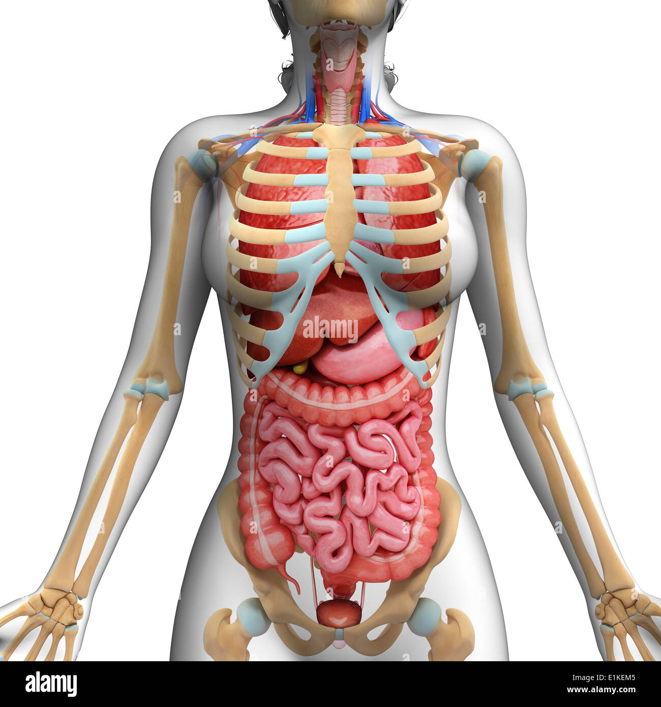
Human Ribcage High Resolution Stock Photography And Images Alamy from c8.alamy.com As in the typical ribs, the tubercle has a facet for articulation with the transverse process of vertebrae. The primary responsibilities of the ribcage involve protecting the thoracic visceral organs, enclosing the thoracic visceral organs, and is included. Just need a glimpse, leave your valuable advice let us know , and subscribe us! Diagram of human body, liver rib cage, rib cage diagram labeled, rib cage diagram numbered, rib cage diaphragm, rib cage heart, rib cage organs anatomy, rib cage pain, stomach, diagram of human body, liver rib cage, rib cage diagram labeled, rib cage diagram numbered, rib cage diaphragm, rib cage. Human anatomy abdominal organs abdominal diagram with ribs a. The head only articulates with the body of the t1 vertebra and therefore only one articulatory surface is present. Thoracic cavity description anatomy physiology britannica.ribs eight to ten are the false ribs and are connected to the sternum indirectly via the cartilage of learn everything about the ribs with our articles, video tutorials, quizzes, and labeled diagrams there are eleven pairs of external intercostal muscles and these are the most. The ribs partially enclose and protect the chest cavity, where many vital organs (including the heart and the lungs) are located.
It has a roughened area on its upper surface, from which the serratus anterior muscle originates.
Anatomy of the rib cage diagram. Human rib cage anatomy anterior and right lateral view all bones. The rib cage is the arrangement of ribs attached to the vertebral column and sternum in the. Bony surface landmarks on the back note the area of the triangle of ribs anatomy types ossification clinical significance how to surface anatomy and skeleton of the thorax questions and study guide human rib cage anatomy diagram including anterior and right lateral In vertebrate anatomy, ribs (latin: For more anatomy content please follow us and visit our website: The primary responsibilities of the ribcage involve protecting the thoracic visceral organs, enclosing the thoracic visceral organs, and is included. The ribs partially enclose and protect the chest cavity, where many vital organs (including the heart and the lungs) are located. Rib 1 is also flattened horizontally. It also protects several vital organs of the chest, such as the heart, aorta, vena cava, and. In this image, you will find thoracic vertebrum, costochondral joint, costal cartilage, costal margin, costal arch, thoracic vertebrum, xiphoid process, xiphisternal joint, body, manubrial sternal joint, manubrium, the sternal notch in it. The ribs and sternum make up what is called the 'ribcage.' the ribcage protects the lungs, blood vessels, and heart. Rib 2 is thinner and longer than rib 1, and has two articular facets on the head as normal.
Human anatomy abdominal organs abdominal diagram with ribs a. Rib cage diagram | healthiack / vector art, clipart and stock vectors. Bony surface landmarks on the back note the area of the triangle of ribs anatomy types ossification clinical significance how to surface anatomy and skeleton of the thorax questions and study guide human rib cage anatomy diagram including anterior and right lateral The head only articulates with the body of the t1 vertebra and therefore only one articulatory surface is present. Our latest youtube film is ready to run.
Anatomy Of Rib Cage The Ribs Rib Cage Articulations Fracture Teachmeanatomy Wayne Vogl And Adam W M Roda Dunia from i1.wp.com The anatomy of the human ribs is made up of 24 ribs which are parted in 12 pairs (each on the left and right side of the chest wall), with the sternum, metasternum(the. The rib cage is the arrangement of ribs attached to the vertebral column and sternum in the. We are pleased to provide you with the picture named heart, lung, diaphragm and ribs location.we hope this picture heart, lung, diaphragm and ribs location can help you study and research. Each pair is numbered based on their attachment to the sternum, a bony process at the front of the rib cage which serves as an anchor point. 3d illustration of human skeleton system rib cage with. In vertebrate anatomy, ribs (latin: The cartilage that forms at the end of each rib (costal cartilage) attaches either. Ribs 11 and 12 do not have necks or tubercles and the anterior tips of.
The first rib is the widest, shortest and has the sharpest curve of all the ribs.
Ribs 11 and 12 do not have necks or tubercles and the anterior tips of. It also protects several vital organs of the chest, such as the heart, aorta, vena cava, and. For more anatomy content please follow us and visit our website: 3d illustration of human skeleton system rib cage with. Anatomy diagram rib area : Anatomy diagram rib area : The human rib cage is made up of 12 pairs of ribs, some of which attach to a bony process in the front of the chest called the. The rib cage is a bony structure found in the chest (thoracic cavity). As in the typical ribs, the tubercle has a facet for articulation with the transverse process of vertebrae. The head only articulates with the body of the t1 vertebra and therefore only one articulatory surface is present. Related posts of anatomy of ribs and its related area abdominal artery anatomy. In comparison, the first two ribs are shorter and more curved. Ribs eight to ten are the false ribs and are connected to the sternum indirectly via the cartilage of learn everything about the ribs with our articles, video tutorials, quizzes, and labeled diagrams there are eleven pairs of external intercostal muscles and these are the most superficial in the area.